Home
/ Anatomy Of Rib Cage Muscles : 1844 Antique ANATOMY print by Lemercier fine lithograph of : The thoracic cage is part of the axial skeleton (also known as the rib cage), and consists of 24 ribs, the sternum, costal cartilage, and the 12 thoracic vertebrae.
Anatomy Of Rib Cage Muscles : 1844 Antique ANATOMY print by Lemercier fine lithograph of : The thoracic cage is part of the axial skeleton (also known as the rib cage), and consists of 24 ribs, the sternum, costal cartilage, and the 12 thoracic vertebrae.
Anatomy Of Rib Cage Muscles : 1844 Antique ANATOMY print by Lemercier fine lithograph of : The thoracic cage is part of the axial skeleton (also known as the rib cage), and consists of 24 ribs, the sternum, costal cartilage, and the 12 thoracic vertebrae.. The thoracic cage is part of the axial skeleton (also known as the rib cage), and consists of 24 ribs, the sternum, costal cartilage, and the 12 thoracic vertebrae. Originate at the lower border of the rib inserting into the superior border of the rib below. Ribs & thoracic cage muscles attachments. In the back, latissimus dorsi and erector spinae muscles (anatomy lesson #10) cover the 11th and 12th ribs of the thoracic cage and deeper yet are the paired abdominal kidneys flanking the. Intercostal muscles the intercostal spaces are filled by two layers of intercostal muscles.
Rendering done with a carestream workstation. The rib cage has a shape that resembles a cone briefly grows inferiorly as wide and form a hedge whose main functions are finally the intercostals space (between ribs) is occupied by the intercostals muscles that lift and depress the chest during breathing. 'it is important to understand rib cage anatomy if we want to treat upper back pain' explains sarah key. The thoracic cage makes up the skeleton for the thoracic wall, and provides the attachments needed for the muscles of the neck, thorax. The thoracic cage (rib cage) is the skeletal framework of the thoracic wall, which encloses the thoracic cavity.
Side Of Rib Cage Muscles : What Causes Pain Under the Left ... from s3.amazonaws.com It provides a strong framework onto which the muscles of the cramps in ribcage are often observed in those who strain or overwork their upper body. During normal breathing, contraction of the major inspiratory muscle, the diaphragm, produces both rib cage expansion and a downward movement of the diaphragm. Rendering done with a carestream workstation. Originate at the lower border of the rib inserting into the superior border of the rib below. The thoracic cage (rib cage) is the skeletal framework of the thoracic wall, which encloses the thoracic cavity. Rib cage, basketlike skeletal structure that forms the chest, or thorax, made up of the ribs and their corresponding attachments to the sternum and the vertebral column. For example, flexor, extensor, adductor and abductor are names associated with the action of the muscle. Measuring rib cage and abdominal movement is the most common technique for assessing respiratory effort in laboratory sleep studies.
Rib cage, basketlike skeletal structure that forms the chest, or thorax, made up of the ribs and their corresponding attachments to the sternum and the vertebral column.
Muscles of thorax, upper extremities, back and diaphragm are given connection by this twelve pairs of ribs are attached to the thoracic vertebrae. During normal breathing, contraction of the major inspiratory muscle, the diaphragm, produces both rib cage expansion and a downward movement of the diaphragm. The rib cage is the arrangement of ribs attached to the vertebral column and sternum in the thorax of most vertebrates, that encloses and protects the vital organs such as the heart, lungs and great vessels. The fibers attach to the rib cage and the pubis of the hip bones. All muscles that are attached to the human rib cage have the. Rendering done with a carestream workstation. In the back, latissimus dorsi and erector spinae muscles (anatomy lesson #10) cover the 11th and 12th ribs of the thoracic cage and deeper yet are the paired abdominal kidneys flanking the. Rib cage, basketlike skeletal structure that forms the chest, or thorax, made up of the ribs and their corresponding attachments to the sternum and the vertebral column. Seventeen muscles attach to the scapula, and it articulates with the clavicle to form the shoulder girdle or pectoral girdle, which. Everyone has nice muscles in ct scanning! The intercostal muscles extend from the vertebrae behind to the sternum in front. Muscles are often named for their primary action. The rib cage surrounds the lungs and the heart, serving as an important means of bony protection for these vital organs.
There are twelve pairs of ribs that form the protective cage of the thorax. Some other muscles that are not part of the thoracic wall but attach to it are the pectoralis major, minor, serratus anterior and the scalene muscles. During normal breathing, contraction of the major inspiratory muscle, the diaphragm, produces both rib cage expansion and a downward movement of the diaphragm. Facets for tubercles of like numbered ribs on its transverse processes. Muscle spasms felt within the rib cage may also be caused by the abdominal muscles.
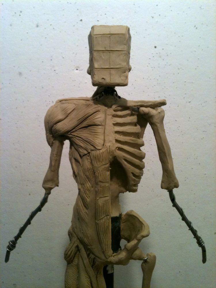
Claire Lordon Design: Écorché Rib Cage to Humerus Bone and ... from 4.bp.blogspot.com The major abdominal muscles include the transverse according to medical news today, it's important to understand the anatomy of the rib cage when determining whether pain under the rib cage is mild. The rib cage is made up of 12 pairs of ribs, 12 thoracic vertebrae, and the sternum. In the back, latissimus dorsi and erector spinae muscles (anatomy lesson #10) cover the 11th and 12th ribs of the thoracic cage and deeper yet are the paired abdominal kidneys flanking the. It provides a strong framework onto which the muscles of the cramps in ribcage are often observed in those who strain or overwork their upper body. Another important feature of the rib cage is the manubriosternal joint also known as the sternal angle of louis. The thoracic cage makes up the skeleton for the thoracic wall, and provides the attachments needed for the muscles of the neck, thorax. See more ideas about anatomy, rib cage anatomy, anatomy study. Rib cage pain can arise from injury to any of the muscles, bones, nerves or joints within the thoracic cage region.
Muscles of thorax, upper extremities, back and diaphragm are given connection by this twelve pairs of ribs are attached to the thoracic vertebrae.
The following general rules regarding actions can be. The other attachment of these muscles is usually considered to be either superior or inferior to the rib attachment. The thoracic cage (rib cage) is the skeletal framework of the thoracic wall, which encloses the thoracic cavity. Learn about ribs muscle with free interactive flashcards. It provides a strong framework onto which the muscles of the cramps in ribcage are often observed in those who strain or overwork their upper body. Another important feature of the rib cage is the manubriosternal joint also known as the sternal angle of louis. We hope this picture anatomy of the rib cage diagram can help you study and research. Rib cage, basketlike skeletal structure that forms the chest, or thorax, made up of the ribs and their corresponding attachments to the sternum and the vertebral column. Rib cage anatomy and its implications in back pain. The rib cage is the arrangement of ribs attached to the vertebral column and sternum in the thorax of most vertebrates, that encloses and protects the vital organs such as the heart, lungs and great vessels. The head of each rib articulates with the costal facet on the body of its own vertebra, and a. Seventeen muscles attach to the scapula, and it articulates with the clavicle to form the shoulder girdle or pectoral girdle, which. Originate at the lower border of the rib inserting into the superior border of the rib below.
Anatomy the rib cage is a bony structure found in the chest thoracic cavity. But for an anatomy study its not. The thoracic cage consists of the 12 thoracic vertebrae, the associated intervertebral discs, 12 pairs of ribs with their costal cartilages, and the sternum. The major abdominal muscles include the transverse according to medical news today, it's important to understand the anatomy of the rib cage when determining whether pain under the rib cage is mild. Rendering done with a carestream workstation.
Pin on skeleton from i.pinimg.com The intercostal muscles extend from the vertebrae behind to the sternum in front. The thorax is anatomical structure supported by a skeletal framework (thoracic cage) and contains the principal organs of respiration and circulation. Some of the most common causes of rib cage pain stem from sports and physical activity. Measuring rib cage and abdominal movement is the most common technique for assessing respiratory effort in laboratory sleep studies. Abdomen & ribs muscle movements. Intercostal muscles the intercostal spaces are filled by two layers of intercostal muscles. Facets for tubercles of like numbered ribs on its transverse processes. The major abdominal muscles include the transverse according to medical news today, it's important to understand the anatomy of the rib cage when determining whether pain under the rib cage is mild.
The rib cage surrounds the lungs and the heart, serving as an important means of bony protection for these vital organs.
Rib cage anatomy and its implications in back pain. But for an anatomy study its not. The intercostal muscles extend from the vertebrae behind to the sternum in front. Seventeen muscles attach to the scapula, and it articulates with the clavicle to form the shoulder girdle or pectoral girdle, which. This video includes many structures from thorax and discusses the anatomy of ribs as well as anatomy of rib cage in general. Originate at the lower border of the rib inserting into the superior border of the rib below. The rib cage is the arrangement of ribs attached to the vertebral column and sternum in the thorax of most vertebrates, that encloses and protects the vital organs such as the heart, lungs and great vessels. 'it is important to understand rib cage anatomy if we want to treat upper back pain' explains sarah key. Thoracic cage is a skeletal framework which supports the thorax. Muscles that move the rib cage attach to the rib cage. During normal breathing, contraction of the major inspiratory muscle, the diaphragm, produces both rib cage expansion and a downward movement of the diaphragm. For more anatomy content please follow us and visit our website: The ribs are curved, flat bones which form the majority of the thoracic cage.
The thoracic cage (rib cage) is the skeletal framework of the thoracic wall, which encloses the thoracic cavity anatomy of rib cage. For example, flexor, extensor, adductor and abductor are names associated with the action of the muscle.


, and consists of 24 ribs, the sternum, costal cartilage, and the 12 thoracic vertebrae.)